Bioclear and Injection Moulding Technique (IMT)

Figure 1: Pre-operative intraoral view. Old restorations,
cracks and discolourations are visible.

Figure 2: After internal bleaching of
tooth #11
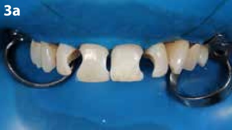
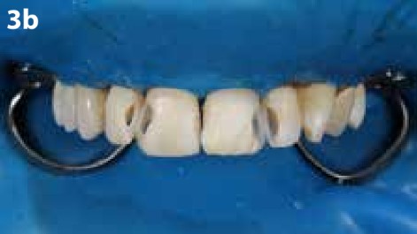
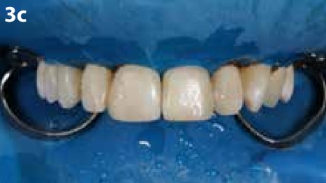
Figure 3: After removal of the old restorations

Figure 4: Smile after replacement of the old
restorations

Figure 5: Wax-up of the
frontal teeth

Figure 6: Transparent mould from EXACLEAR

Figure 7: Creation of the injection channels
with the tip of the syringe

Figure 8: Frontal teeth were cleaned and
slightly roughened

Figure 9: Frontal teeth were etched with
phosphoric acid

Figure 10: Frosty appearance of the teeth
after etching

Figure 11: Teflon tape was applied on the
adjacent teeth
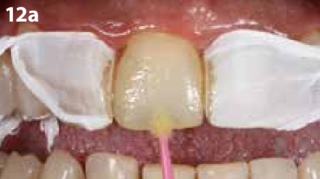
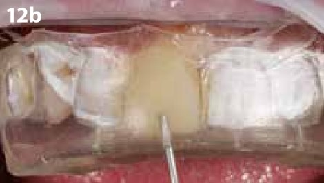
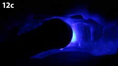
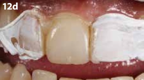
Figure 12: a) Bonding with G-Premio BOND; b) Injection of G-ænial Universal Injectable (Shade A2); c) Light-curing through the EXACLEAR mould d) After removal of the mould. Excess could be easily removed.

Figure 13: Intraoral view before polishing
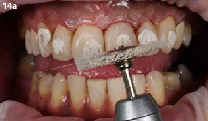
Figure 14a: Polishing with soft brushes
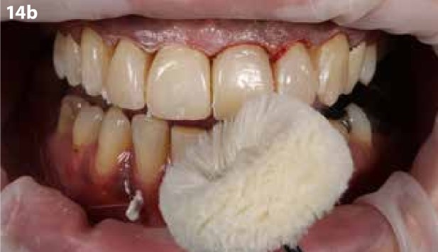
Figure 14a: Polishing with soft brushes
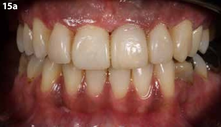
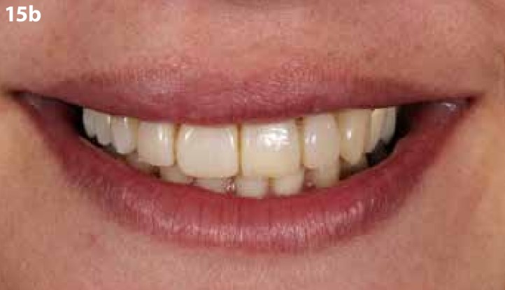
Figure 15: After treatment. a) Intraoral view; b) Smile

